Bioimaging Research Group
Research Outline
Developing Imaging and Sensing Technologies to Support the Future of the Bioindustry
Various targets exist all around us, including biomolecules and biological tissues, as well as food products and environmental substances. These targets vary widely in size and concentration, making the development of new technologies essential for accurately “visualizing” and “measuring” them. Our group is engaged in the research and development of highly original observation and measurement technologies, devices, materials, and methodologies aimed at realizing next-generation imaging and sensing technologies. In addition, we actively promote the practical application and social implementation of our research outcomes to contribute to society.
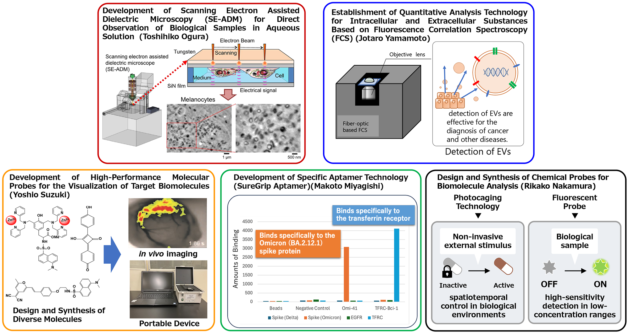
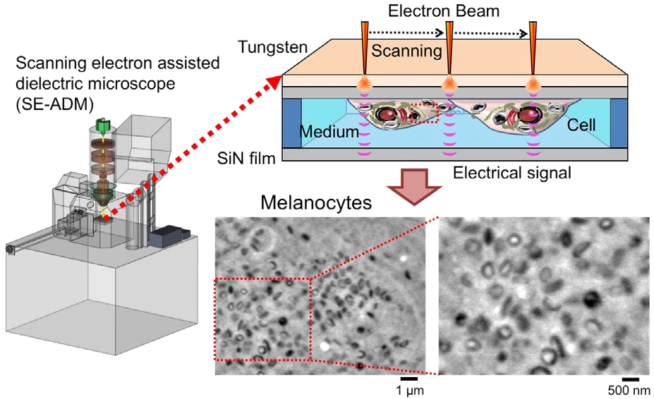
Development of Scanning Electron Assisted Dielectric Microscopy (SE-ADM) for Direct Observation of Biological Samples in Aqueous Solution (Toshihiko Ogura)
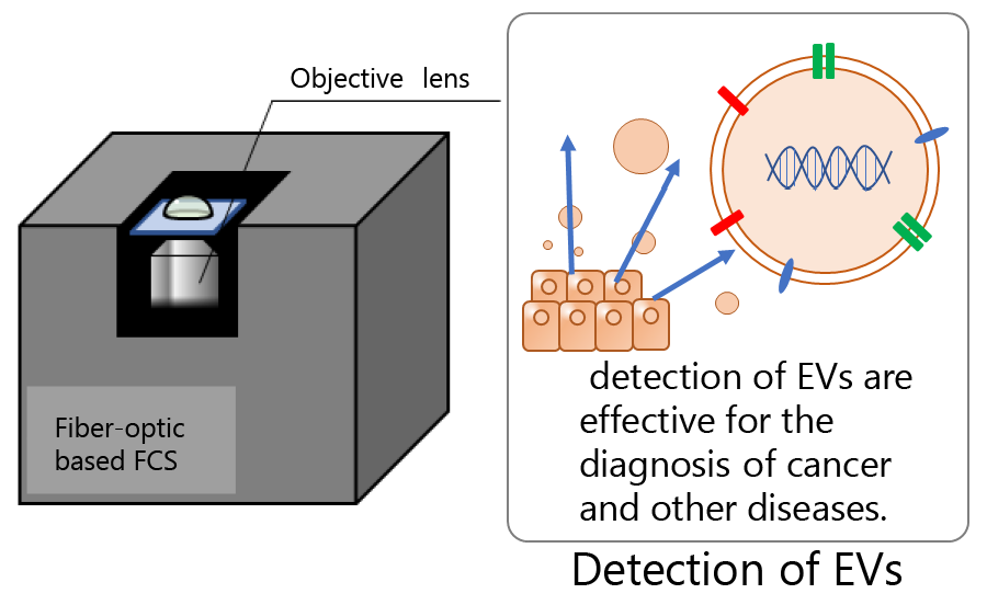
Establishment of Quantitative Analysis Technology for Intracellular and Extracellular Substances Based on Fluorescence Correlation Spectroscopy (FCS) (Jotaro Yamamoto)
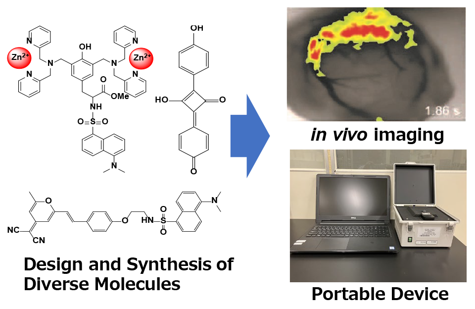
Development of High-Performance Molecular Probes for the Visualization of Target Biomolecules (Yoshio Suzuki)
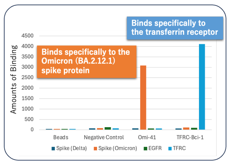
Development of Specific Aptamer Technology (SureGrip Aptamer)(Makoto Miyagishi)
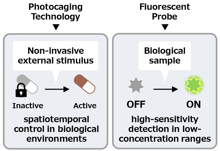
Design and Synthesis of Chemical Probes for Biomolecule Analysis (Rikako Nakamura)