Visualization of Spatial Partitioning of Glycans in Cardiac Fibrotic Tissue
A research team at AIST, including Patcharaporn Boottanun (Invited Collaborative Researcher), Chiaki Nagai-Okatani (Group Leader), and Atsushi Kuno (Principal Researcher), in collaboration with Prof. Kunio Kawanishi and Assistant Prof. Masaki Baba of Showa Medical University (also affiliated with the University of Tsukuba), revealed that glycans are spatially partitioned across distinct pathological microenvironments in myocardial fibrotic tissue.
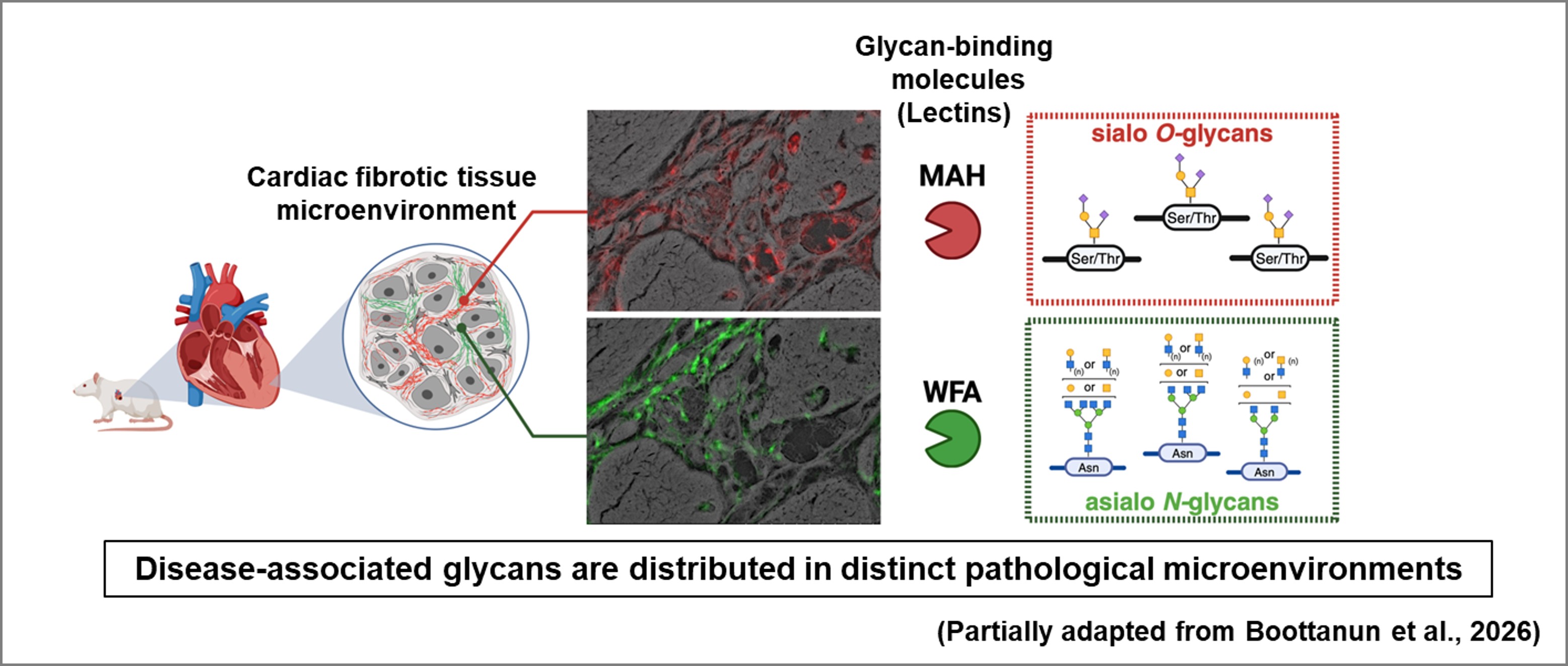
In this study, the team applied a lectin microarray–based tissue glycome profiling technology developed at AIST [1,2] to a hypertensive heart failure model with myocardial fibrosis, successfully capturing glycan alterations associated with fibrosis. However, this approach alone had limitations in determining the precise spatial localization of these glycans within tissue structures. To overcome this limitation, they combined this approach with a high-resolution imaging method developed by Kawanishi and Baba, which captures fine tissue microstructures including depth (Z-axis) information. This integration enabled visualization of glycan distribution in direct correspondence with tissue architecture. As a result, sialylated O-glycans and non-sialylated N-glycans were found to be selectively localized in distinct pathological microenvironments.
This technology enables the analysis of disease microenvironments using formalin-fixed paraffin-embedded (FFPE) tissue specimens accumulated in clinical settings and is expected to facilitate drug discovery by identifying aberrantly glycosylated proteins as potential therapeutic targets.
Spatial distribution of glycans in cardiac fibrotic tissue
Publication
- Title: : A hierarchical lectin-based multimodal workflow for spatial mapping of extracellular matrix glycans in fibrotic hearts
- Authors: : Patcharaporn Boottanun, Chiaki Nagai-Okatani*, Kunio Kawanishi, Masaki Baba, Tomofumi Nakatsukasa, Tomoko Ishizu, Kiyohiko Angata, Atsushi Kuno
- Journal:Analytical and Bioanalytical Chemistry (published online: April 27, 2026)
- DOI: 10.1007/s00216-026-06501-6
Related articles
[1] Zou X et al. A standardized method for lectin microarray-based tissue glycome mapping. Sci Rep, 7:43560, 2017. doi:10.1038/srep43560
[2] Boottanun P et al. An improved evanescent fluorescence scanner suitable for high-resolution glycome mapping of formalin-fixed paraffin-embedded tissue sections. Anal Bioanal Chem, 415(28):6975–6984, 2023. doi:10.1007/s00216-023-04824-2