Multimodal Spatial Molecular Imaging Research Group
Multimodal Spatial Molecular Imaging Research Group Overview
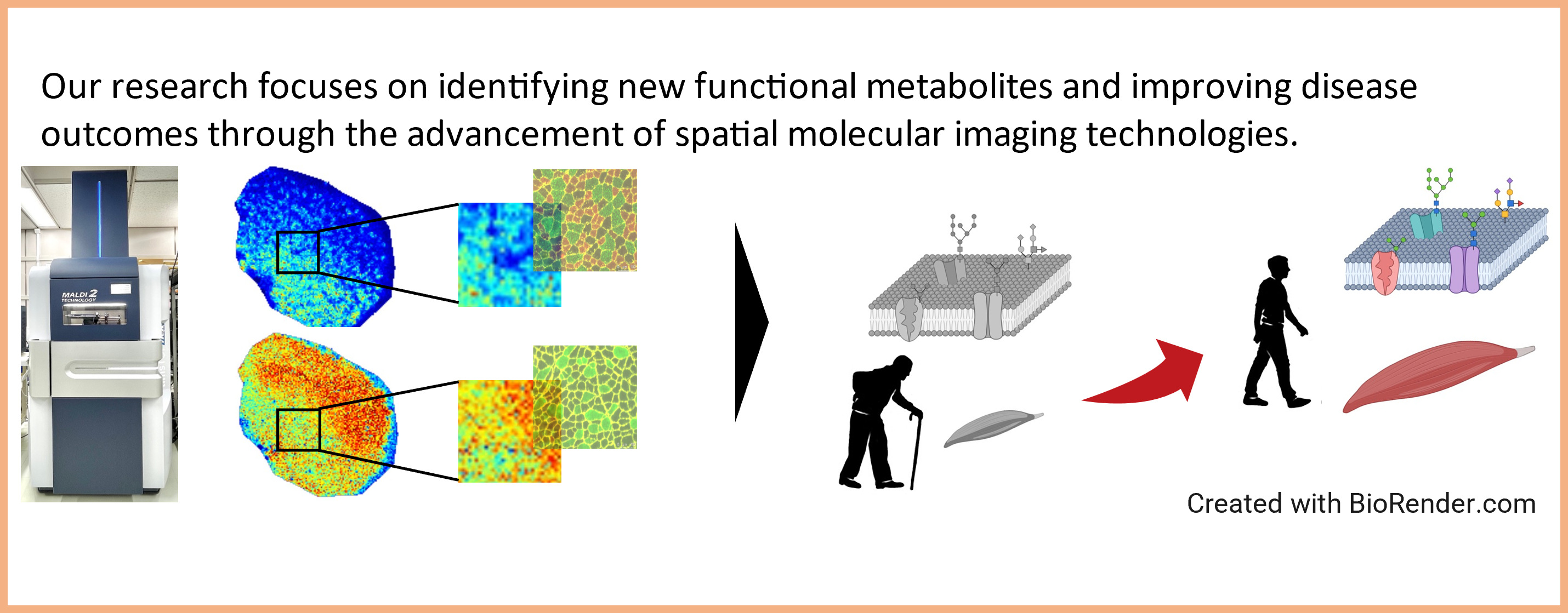
The main goal of this research group is to develop imaging techniques to visualize the spatial expression information of functional molecules (such as glycans and proteins) in tissues and cells using mass spectrometry and microscopy. In addition, we are developing technologies for the evaluation and control of cellular states and functions using cell models such as cultured cells, stem cell-derived cells, and organoids. By visualizing health conditions at the molecular level and accelerating the development of diagnostic and therapeutic methods for diseases based on an understanding of the molecular basis of life phenomena, we aim to contribute to the realization of a healthy and long-lived society.
Research Project
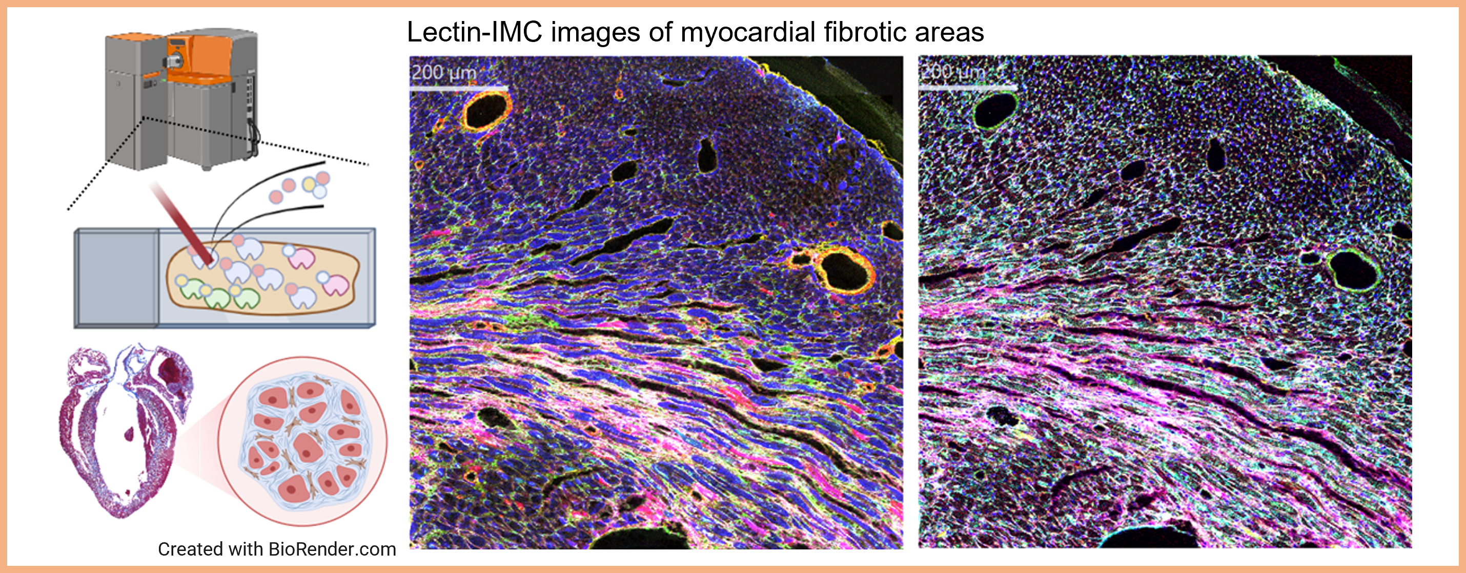
Project 1:Development of multiplexed spatial analysis technology to “visualize” glycans on tissue sections
Researcher: NAGAI-OKATANI Chiaki
For the development of “glyco-medicines” based on disease-related changes in protein glycosylation, it is important to identify glycoproteins (i.e., the combinations of glycans and proteins) that are truly associated with pathological conditions and highly specific to diseased cells. As a method to evaluate disease-related glycan and glycoprotein candidates obtained by glyco-multiomics in conjunction with pathological observations, we have established a spatial analysis method using lectin-assisted imaging mass cytometry (Lectin-IMC) to simultaneously detect glycans and proteins on tissue samples at the single cell level. This method allows the efficient selection of lectins that recognize disease-related glycans and verification of candidate glycoproteins identified by glycoproteome analysis in relation to pathological conditions, which is expected to accelerate the identification of promising seeds for drug discovery. This technology will also be useful for elucidating glycan functions in intercellular communication.
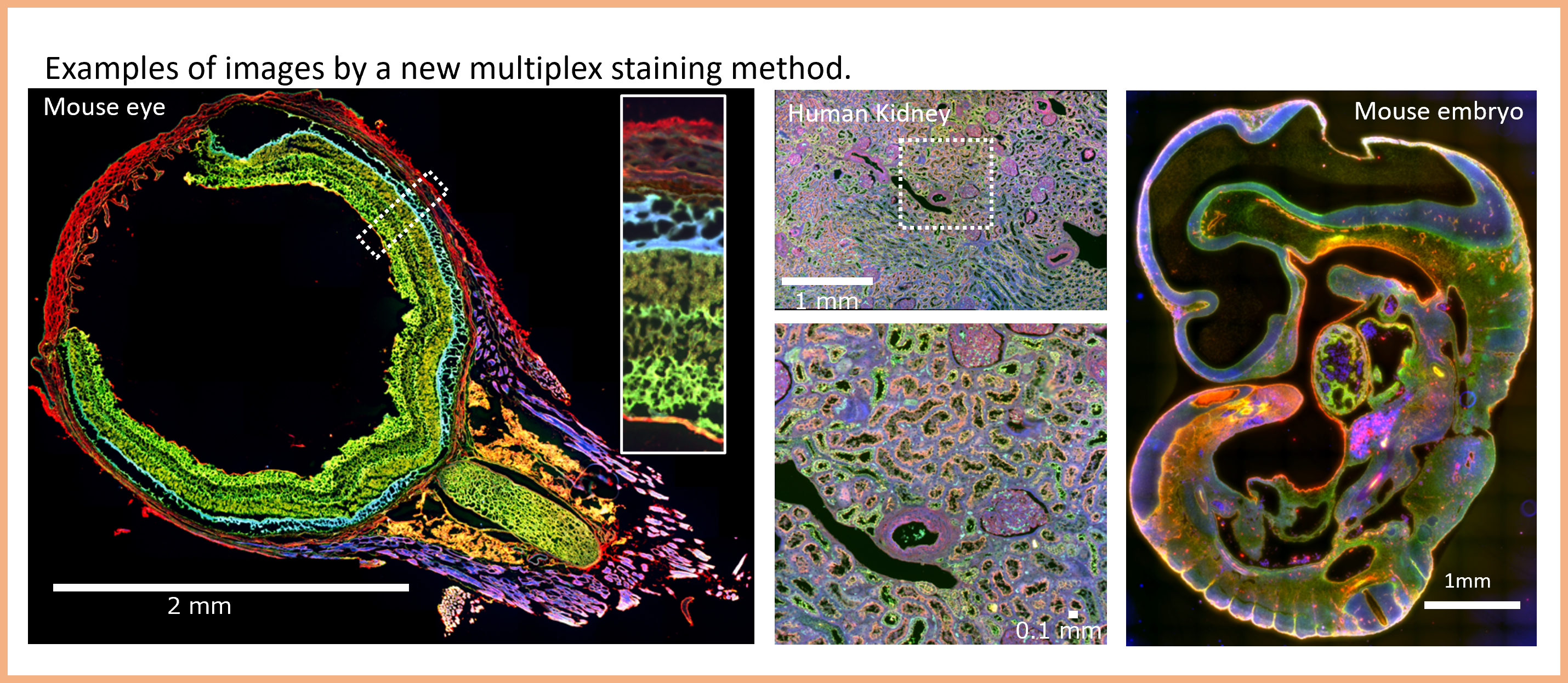
Project 2:Development of a new diagnostic technology using multiplex staining
Researcher: NAGASAKI Akira
Even though tens of millions of pathological diagnoses are performed annually, there are only about 2000 pathologists in charge of these diagnoses, leading to a chronic shortage of specialists. In addition, pathological diagnosis relies on specialized skills such as techniques and experience. To solve this social issue, the digitization of histopathology images and remote diagnosis via networks are promoted, and AI-based diagnosis is also attempted for practical use. Therefore, this study aims to develop a new multiplex fluorescence imaging methods specifically designed for AI-based diagnosis.
Project 3:Development of Cell Evaluation and Control Technologies for Understanding and Preventing Age-Related Diseases
Researcher: AKAGI Yuka
As society ages, the increasing prevalence of age-related diseases, including neurodegenerative disorders, has become a major challenge for extending healthy life expectancy and reducing medical and nursing care burdens. My research focuses on developing technologies to visualize and evaluate changes in cellular states and functions associated with aging and disease, using cellular models such as cultured cells, stem cell-derived cells, and organoids, while minimizing damage to the cells. By focusing on how cells sense and respond to their surrounding environment, including glycans, inter-organ communication, and physical stimuli, I aim to understand cellular changes involved in disease onset and progression. Ultimately, this research seeks to contribute to the prevention, diagnosis, drug discovery, and cell manufacturing technologies for age-related diseases.
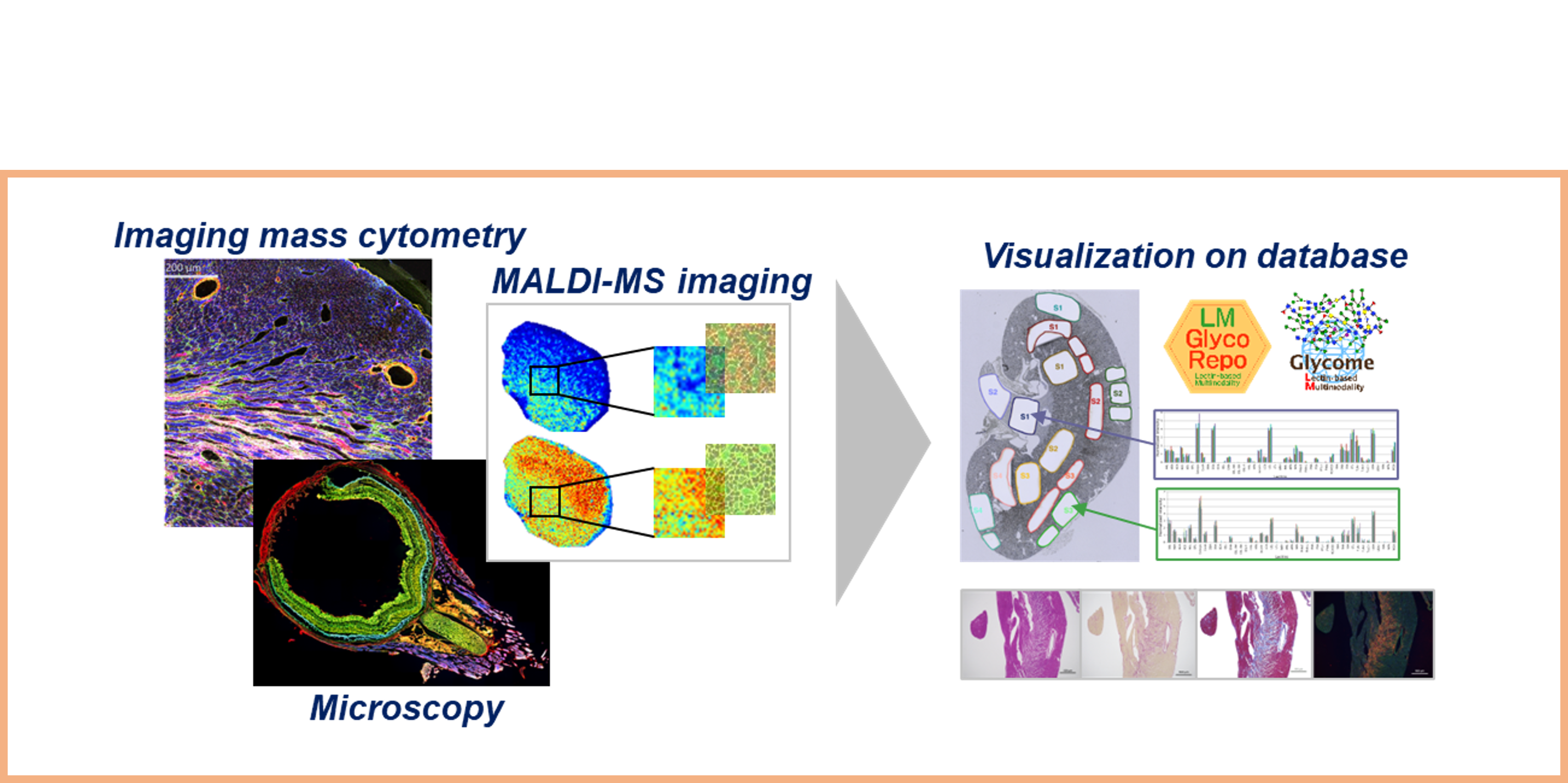
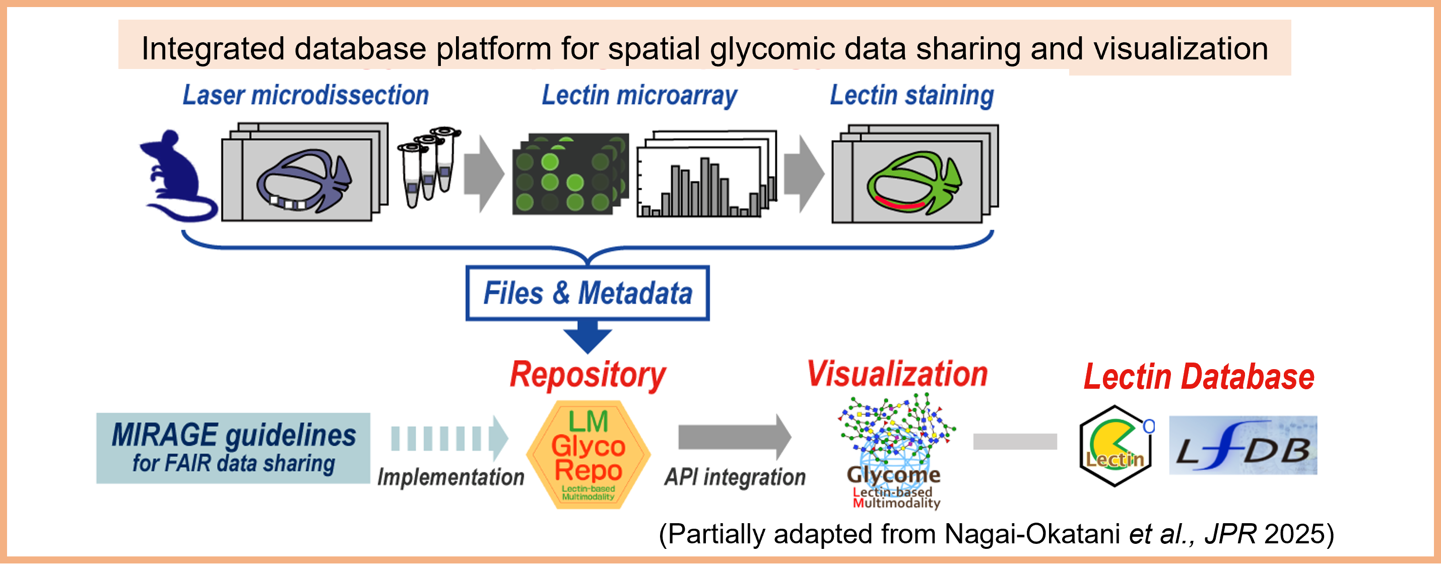
Project 4:Development of an Integrated Database Infrastructure for Glycan and Spatial Molecular Information
Researcher: NAGAI-OKATANI Chiaki
We are developing a database infrastructure that enables the integrated management and visualization of diverse spatial molecular imaging data linked with glycan information. In collaboration with the Molecular and Cellular Glycoproteomics Research Group, we aim to establish an environment for cross-sectional analysis of tissue-, cell-, and molecule-level information by integrating glycan-related resources, including lectin-related repositories and databases such as LfDB, LM-GlycoRepo, and LM-GlycomeAtlas. Through this effort, we seek to advance the discovery of disease-associated molecules and enhance the utilization and value of glycan-related data.
Staff Members
| photo | position & name | field of expertise | and other info |
|---|---|---|---|
 |
Research Group Leader NAGAI-OKATANI Chiaki |
|
|
 |
Senior Researcher NAGASAKI Akira |
|
|
|
Senior Researcher AKAGI Yuka |
|
|
|
Researcher MIYAKO Kei |
|
|
Publications
- Boottanun, P; Nagai-Okatani, C; Kawanishi, K; Baba, M; Nakatsukasa, T; Ishizu, T; Angata, K; Kuno, A.
A hierarchical lectin-based multimodal workflow for spatial mapping of extracellular matrix glycans in fibrotic hearts.
Analytical and bioanalytical chemistry. 2026 Apr 27. doi:10.1007/s00216-026-06501-6 - Boottanun, P; Fuseya, S; Kuno, A; Nagai-Okatani, C.
From Laser Microdissection to Spatial Glycomics: Lectin Microarray Protocols for Tissue Glycome Mapping with High-End Scanners.
METHODS MOL BIOL. 2026;3015:21-41. doi:10.1007/978-1-0716-5154-4_3 - Kano, R; Kusano, T; Ando, T; Takeda, R; Natsuyama, R; Takagi, R; Poole, DC; Kano, Y; Hoshino, D.
Effects of local skeletal muscle cooling prior to acute exercise and endurance training on mitochondrial adaptation in mice.
JOURNAL OF PHYSIOLOGICAL SCIENCES. 2026 Mar;76(1):100059. doi:10.1016/j.jphyss.2026.100059 - Nagai-Okatani, C; Hosoda, M; Hsiung, W; Sato, T; Sakaue, H; Fuseya, S; Fukuda, E; Yamada, I; Aoki-Kinoshita, KF; Kuno, A.
Meeting Report on the 1st GlyCosmos Symposium.
GLYCOBIOLOGY. 2025 Nov 21:cwaf079. doi:10.1093/glycob/cwaf079 - Dang, Cao TL; Kawanishi, K; Hashimoto, S; Hengphasatporn, K; Nagai-Okatani, C; imura, T; Abdelaziz, M; Shiratani, R; Poullikkas, T; Azmi, NU; Baba, M; Okita, Y; Watanabe, Y; Bando, H; Yamazaki, S; Shigeta, Y; Kuno, A; SKato, M.
Tumor-expressed GPNMB orchestrates Siglec-9(+) TAM polarization and EMT to promote metastasis in triple-negative breast cancer.
PROC NATL ACAD SCI U S A. 2025 Sep 9;122(36):e2503081122. doi: 10.1073/pnas.2503081122 - Ando, T; Takeda, R; Kano, R; Kusano, T; Nonaka, Y; Kano, Y; Hoshino, D.
Effects of pyruvate administration on mRNA expression of inflammatory cytokines in adipose tissue and whole-body glucose metabolism in male mice.
PHYSIOL REP. 2025 Aug;13(15):e70362. doi:10.14814/phy2.70362 - Nagai-Okatani, C; Fujita, N; Boottanun, P; Tanaka, M; Shiota, M; Shinmachi, D; Angata, K; Aoki-Kinoshita, K; Kuno, A.
LM-GlycoRepo Version 1.0: A Novel Repository System for Mouse Tissue Glycome Mapping Data.
JOURNAL OF PROTEOME RESEARCH. 2025 JUN 11. doi:10.1021/acs.jproteome.5c00184 - Kano, R; Takeda, R; Sotani, Y; Takagi, R; Tabuchi, A; Shirakawa, H; Poole, DC; Kano, Y; Hoshino, D.
Cooling-induced changes in intracellular hydrogen peroxide and gene expression in mouse skeletal muscle in vivo.
AM J PHYSIOL REGUL INTEGR COMP PHYSIOL. 2025 May 7. doi:10.1152/ajpregu.00014.2025 - Nagai-Okatani, C; Tomioka, A; Tominaga, D; Sakaue, H; Kuno, A; Kaji, H.
Inter-tissue glycan heterogeneity: site-specific glycoform analysis of mouse tissue N-glycoproteomes using MS1-based glycopeptide detection method assisted by lectin microarray.
ANALYTICAL AND BIOANALYTICAL CHEMISTRY. 2024 Dec 16. doi:10.1007/s00216-024-05686-y - Kudo, K; Nishimura, T; Miyako, K; Suenaga, H; Shin-Ya, K.
A new cannabigerolic acid derivative and its unprenylated precursor produced through the reconstitution of cannabinoid biosynthesis in Streptomyces.
J ANTIBIOT (Tokyo). 2024 Dec 3. doi:10.1038/s41429-024-00793-5 - Nagai-Okatani, C; Tominaga, D; Tomioka, A; Sakaue, H; Goda, N; Ko, S; Kuno, A; Kaji, H.
GRable version 1.0: A software tool for site-specific glycoform analysis with improved MS1-based glycopeptide detection with parallel clustering and confidence evaluation with MS2 information.
MOL CELL PROTEOMICS. 2024 Aug 22:100833. doi: 10.1016/j.mcpro.2024.100833 - Kano, R; Kusano, T; Takeda, R; Shirakawa, H; Poole, DC; Kano, Y; Hoshino, D.
Eccentric contraction increases hydrogen peroxide levels and alters gene expression through Nox2 in skeletal muscle of male mice.
J APPL PHYSIOL (1985). 2024 Jul 25. doi: 10.1152/japplphysiol.00335.2024 - Akagi, Y; Takayama, Y; Nihashi, Y; Yamashita, A; Yoshida, R; Miyamoto, Y; Kida, YS.
Functional engineering of human iPSC-derived parasympathetic neurons enhances responsiveness to gastrointestinal hormones.
FEBS OPEN BIO. 2023 Nov 27. doi: 10.1002/2211-5463.13741 - Boottanun, P; Nagai-Okatani, C; Nagai, M; Ungkulpasvich, U; Yamane, S; Yamada, M; Kuno, A.
An improved evanescent fluorescence scanner suitable for high-resolution glycome mapping of formalin-fixed paraffin-embedded tissue sections.
ANALYTICAL AND BIOANALYTICAL CHEMISTRY. 2023 JUL 3. doi: 10.1007/s00216-023-04824-2 - Takayama, Y, Akagi, Y, Kida, YS.
Deciphering the Molecular Mechanisms of Autonomic Nervous System Neuron Induction through Integrative Bioinformatics Analysis.
INT J MOL SCI. 2023 May 21;24(10):9053. doi: 10.3390/ijms24109053 - Sogo, T; Nakao, S; Tsukamoto, T; Ueyama, T; Harada, Y; Ihara, D; Ishida, T; Nakahara, M; Hasegawa, K; Akagi, Y; Kida, YS; Nakagawa, O; Nagamune, T; Kawahara, M; Kawamura, T.
Canonical Wnt signaling activation by chimeric antigen receptors for efficient cardiac differentiation from mouse embryonic stem cells.
INFLAMM REGEN. 2023 Feb 10;43(1):11. doi: 10.1186/s41232-023-00258-6 - Itakura, Y; Hasegawa, Y; Kikkawa, Y; Murakami, Y; Sugiura, K; Nagai-Okatani, C; Sasaki, N; Umemura, M; Takahashi, Y; Kimura, T; Kuno, A; Ishiwata, T; Toyoda, M.
Spatiotemporal changes of tissue glycans depending on localization in cardiac aging.
REGEN THER. 2023 Jan 7;22:68-78. doi: 10.1016/j.reth.2022.12.009 - Miyazaki, Y; Mori, N; Akagi, Y; Oda, T; Kida, YS.
Potential Metabolite Markers for Pancreatic Cancer Identified by Metabolomic Analysis of Induced Cancer-Associated Fibroblasts.
CANCERS. 2022 Mar 8;14(6):1375. doi: 10.3390/cancers14061375 - Nagai-Okatani, C; Zou, X; Matsuda, A; Itakura, Y; Toyoda, M; Zhang, Y; Kuno, A.
Tissue Glycome Mapping: Lectin Microarray-Based Differential Glycomic Analysis of Formalin-Fixed Paraffin-Embedded Tissue Sections.
METHODS MOL BIOL. 2022;2460:161-180. doi: 10.1007/978-1-0716-2148-6_10 - Akagi, Y; Mori, N; Kawamura, T; Takayama, Y; Kida, YS.
Non-invasive cell classification using the Paint Raman Express Spectroscopy System (PRESS)
SCI REP. 2021 Apr 23;11(1):8818. doi: 10.1038/s41598-021-88056-3